My Sick Liver
Liver Cancer
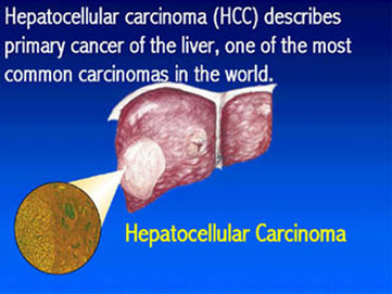
Liver cancer (HepatoCellular Carcinoma or HCC) is a cancer arising from the liver. It is also known as primary liver cancer or hepatoma. The liver is made up of different cell types: liver cells (80%), bile ducts, blood vessels and fat-storing cells. The majority of primary liver cancers (over 90%-95%) arises from liver cells.
When patients or physicians speak of liver cancer they are often referring to cancer that has spread to the liver, having originated in other organs such as the colon, stomach, pancreas, breast and lung. More specifically, this type of liver cancer is called metastatic liver cancer or secondary liver cancer. This is a much more common problem around the world than primary liver cancer and frequently leads to confusion, because the term liver cancer actually can refer to either metastatic liver cancer or hepatocellular cancer. The subject of this page is hepatocellular carcinoma, herein referred to as just liver cancer.
Liver cancer is the third most common cancer in the world. A deadly cancer, liver cancer will kill almost all patients who have it within a year. In 2000, it was estimated that there were about 564,000 new cases of liver cancer worldwide, and a similar number of patients died as a result of this disease. About three-quarters of the cases of liver cancer are found in Southeast Asia. Liver cancer is also very common in sub-Saharan Africa.
What are liver cancer causes and risk factors?
Hepatitis B
A hepatitis B infection can be caught from contaminated blood products, used needles or sexual contact but is frequent among Asian children from contamination at birth or even biting among children at play. The role of hepatitis B virus (HBV) infection in causing liver cancer is well established. The patients with HBV who are at greatest risk for liver cancer are men who also have a family history of liver cancer.
Hepatitis C
Hepatitis C virus (HCV) infection is more difficult to get than hepatitis B. It usually requires direct contact with infected blood, from either contaminated blood products or needles. HCV is also associated with the development of liver cancer. The majority of HCV patients with liver cancer have associated cirrhosis. In several studies of the natural history of hepatitis C, the average time to develop liver cancer after exposure to HCV was about 28 years. In patients that have HCV cirrhosis liver cancer occurred about eight to 10 years after the development of cirrhosis.
In HCV patients, the risk factors for developing liver cancer include the presence of cirrhosis, older age, male gender, elevated baseline alpha-fetoprotein level (AFP), alcohol use and co-infection with HBV.
Alcohol
Cirrhosis caused by chronic alcohol consumption is the most common association of liver cancer in the developed world. In fact, at autopsy, as many as half of alcoholics previously unsuspected to have cancer will have early evidence of cancer hidden within the liver. Many of these people are also infected with chronic HCV. The usual setting is an individual with alcoholic cirrhosis who has stopped drinking for 10 years and then develops liver cancer. It is somewhat unusual for an actively drinking alcoholic to develop liver cancer. What happens is that when the drinking is stopped, the liver cells try to heal by regenerating. It is during this active regeneration that a cancer-producing genetic change can occur, which explains the occurrence of liver cancer after the drinking has been stopped.
More importantly, if an alcoholic does not stop drinking, he or she is unlikely to live long enough to develop the cancer. Alcoholics who are actively drinking are more likely to die from non-cancer related complications of alcoholic liver disease (for example, liver failure). Indeed, patients with alcoholic cirrhosis who die of liver cancer are about 10 years older than patients who die of non-cancer causes. Alcohol adds to the risk of developing liver cancer in patients with chronic HCV or HBV infections.
Aflatoxin B1
Aflatoxin B1 is the most potent liver cancer-forming chemical known. It is a product of a mold called Aspergillus flavus, which is found in food that has been stored in a hot and humid environment. This mold is found in such foods as peanuts, rice, soybeans, corn and wheat. This mold has also been found in marijuana. Aflatoxin B1 is thought to cause cancer by producing changes in the p53 gene. These mutations work by interfering with the gene's important tumor suppressing functions.
Drugs, medications and chemicals
There are no medications that cause liver cancer, but female hormones (estrogens) and protein-building (anabolic) steroids are associated with the development of hepatic adenomas. These are benign liver tumors that may have the potential to become malignant. Thus, in some individuals, hepatic adenoma can evolve into cancer.
Hemochromatosis
Liver cancer will develop in up to 30% of patients with hereditary hemochromatosis. Patients at the greatest risk are those who develop cirrhosis with their hemochromatosis. Unfortunately, once cirrhosis is established, effective removal of excess iron will not reduce the risk of developing liver cancer.
Diabetes and obesity
Over the past decade, the incidence of liver cancer in the United States has risen significantly, paralleling the rise in obesity. Although it is hard to separate the effects of diabetes from obesity on the liver, both conditions can cause chronic damage and accumulation of fat within the liver. This is a disease called NASH (non-alcoholic steatohepatitis), which is present in up to 5% of North Americans. Fatty liver disease like this causes damage to the individual liver cells and may lead to cirrhosis in some people, thereby increasing the risk of liver cancer. Not only is the chance of developing the cancer enhanced, but also patients with diabetes who undergo surgical removal of liver cancer have a higher chance of the cancer returning than do those without diabetes.
Cirrhosis
Individuals with most types of cirrhosis of the liver are at an increased risk of developing liver cancer. In addition to the conditions described above (hepatitis B, hepatitis C, alcohol, and hemochromatosis), alpha 1 anti-trypsin deficiency, a hereditary condition that can cause emphysema and cirrhosis, may lead to liver cancer. Liver cancer is also strongly associated with hereditary tyrosinemia, a childhood biochemical abnormality that results in early cirrhosis.
Certain causes of cirrhosis are less frequently associated with liver cancer than are other causes. For example, liver cancer is rarely seen with the cirrhosis in Wilson's disease or primary sclerosing cholangitis (PSC). It used to be thought that liver cancer is rarely found in primary biliary cirrhosis (PBC) as well. Recent studies, however, show that the frequency of liver cancer in PBC is comparable to that in other forms of cirrhosis.
What are liver cancer symptoms and signs?
The initial symptoms of liver cancer are variable. It is becoming much more common for patients to be identified by screening people at high risk for the cancer and finding the cancer before there are any symptoms at all. There are no specific symptoms of liver cancer, and in fact, the earliest signs are usually subtle and can be mistaken for simple worsening of cirrhosis and liver function. Abdominal pain is uncommon with liver cancer and usually signifies a very large tumor or widespread involvement of the liver. Unexplained weight loss or unexplained fevers are warning signs of liver cancer in patients with cirrhosis.
A common initial presentation of liver cancer in a patient with compensated cirrhosis is the sudden onset of a complication. For example, the sudden appearance of ascites, jaundice or muscle wasting without causative factors suggests the possibility of liver cancer. Cancer can invade and block the portal vein. When this happens, the blood will travel paths of less resistance, such as through esophageal veins. This causes increased pressure in these veins, which results in dilated veins called esophageal varices. The patient then is at risk for hemorrhage from the rupture of the varices into the gastrointestinal tract. Rarely, the cancer itself can rupture and bleed into the abdominal cavity, resulting in bloody ascites.
On physical examination, an enlarged, sometimes tender, liver is the most common finding. Liver cancers are very vascular tumors. Increased amounts of blood feed into the hepatic artery and cause turbulent blood flow in the artery. The turbulence results in a distinct sound in the liver that can be heard with a stethoscope in about one-quarter to one-half of patients with liver cancer. Any sign of advanced liver disease means a poor prognosis. Rarely, a patient with liver cancer can become suddenly jaundiced when the tumor erodes into the bile duct. The jaundice occurs in this situation because both sloughing of the tumor into the duct and bleeding that clots in the duct can block the duct.
In advanced liver cancer, the tumor can metastasize to neighboring tissues or, through the blood vessels, elsewhere in the body. Liver cancer can invade the hepatic veins that drain the liver then blocking these veins, resulting in congestion of the liver. The congestion occurs because the blocked veins cannot drain the blood out of the liver. Normally, the blood in the hepatic veins leaving the liver flows through the inferior vena cava, which is the largest vein that drains into the heart. Blockage of either the hepatic veins or the inferior vena cava results in a very swollen liver and massive formation of ascites.
Liver cancer frequently spreads to the lungs, presumably by way of the bloodstream. Usually, patients do not have symptoms from the lung metastases, which are diagnosed by radiologic studies. Rarely, in very advanced cases, liver cancer can spread to the bone or brain. These are an infrequent problem in many patients who do not live long enough to develop these complications.
How is liver cancer diagnosed?
Blood tests
Liver cancer is not diagnosed by routine blood tests, including a standard panel of liver tests. This is why the diagnosis of liver cancer depends so much on the vigilance of the physician screening with a tumor marker in the blood and radiological imaging studies. Since most patients with liver cancer have associated liver disease, their liver blood tests may not be normal to begin with. If these blood tests become abnormal or worsen due to liver cancer, this usually signifies extensive cancerous involvement of the liver. At that time, any medical or surgical treatment may be too late.
Some abnormal blood tests can indicate the presence of liver cancer. Remember that each cell type in the body contains the full complement of genetic information. What differentiates one cell type from another is the particular set of genes that are turned on or off in that cell. When cells become cancerous, certain of the cell's genes that were turned off may become turned on. Thus, in liver cancer, the cancerous liver cells may take on the characteristics of other types of cells. For example, liver cancer cells sometimes can produce hormones that are ordinarily produced in other body systems. These hormones then can cause certain abnormal blood tests, such as a high red blood count, low blood sugar and high blood calcium.
There is no reliable or accurate screening blood test for liver cancer. The most widely used biochemical blood test is alpha-fetoprotein (AFP), which is a protein normally made by the immature liver cells in the fetus. At birth, infants have relatively high levels of AFP, which fall to normal adult levels by the first year of life.
In adults, high blood levels (over 500 nanograms/milliliter) of AFP are seen in only three situations:
- Liver cancer
- Germ cell tumors (cancer of the testes and ovaries)
- Metastatic cancer in the liver (originating in other organs)
An elevated AFP blood test is seen in about 60% of liver cancer patients. That leaves 40% of patients with liver cancer who have normal AFP levels. Therefore, a normal AFP does not exclude liver cancer. An abnormal AFP does not mean that a patient has liver cancer. It is important to note that patients with cirrhosis and an abnormal AFP, despite having no documentable liver cancer, still are at very high risk of developing liver cancer. Thus, any patient with cirrhosis and an elevated AFP, particularly with steadily rising blood levels, will either most likely develop liver cancer or actually already have an undiscovered liver cancer.
An AFP greater than 500 ng/ml is very suggestive of liver cancer. In fact, the blood level of AFP loosely relates to the aggressiveness of the liver cancer. In patients with liver cancer and abnormal AFP levels, the AFP may be used as a marker of response to treatment. For example, an elevated AFP is expected to fall to normal in a patient whose liver cancer is successfully removed surgically (resected). People with higher AFP levels generally do not live as long as those with lower AFP levels.
Imaging studies
Imaging studies play a very important role in the diagnosis of liver cancer. A good study can provide information as to the size of the tumor, the number of tumors, and whether the tumor has involved major blood vessels locally or spread outside of the liver. There are several types of studies, each having its merits and disadvantages. In practice, several studies combined often complement each other. On the other hand, a plain X-ray is not very helpful, and therefore, is not routinely done in the diagnostic work-up of liver cancer. Further, there is no practical role for nuclear medicine scans of the liver and spleen in the workup for liver cancer. Such scans are not very sensitive and they provide no additional information beyond that provided by the other (ultrasound, CT and MRI) scans.
Advances in ultrasound, CT and MRI technology have almost eliminated the need for angiography. An angiography procedure involves inserting a catheter into the femoral artery through to the aorta, and then into the hepatic artery, the artery that supplies blood to the liver. Contrast material is then injected, and X-ray pictures of the arterial blood supply to the liver are taken. An angiogram of liver cancer shows a characteristic blush that is produced by newly formed abnormal small arteries that feed the tumor.
Another potential test used for many other cancers is a PET (positron emission tomography) scan, which involves the injection of radioactive sugar to light up actively growing cells, as in cancers. However, this is not very useful in liver cancer.
What is the best imaging study for diagnosing liver cancer?
There is no simple answer. Many factors need to be taken into consideration. For example, is the diagnosis of liver cancer known or is the scan being done for screening? What is the expertise of doctors in the patient's area? What is the quality of the different scanners at a particular facility? Are there economic considerations? Does the patient have any other conditions that need to be considered, such as claustrophobia or kidney impairment? Does the patient have any hardware, for example, a pacemaker or metal prosthetic device? (The hardware would make doing an MRI impossible.)
UltraSound
An US examination is usually the first study ordered if liver cancer is suspected in a patient. The accuracy of an ultrasound depends very much on the technician and radiologist who perform the study. Studies report that ultrasound is the most sensitive imaging study for diagnosing and characterizing liver cancer. But in these studies, highly experienced individuals performed the scans and spent up to one hour scanning each patient suspected of having liver cancer. An ultrasound has the advantages of not requiring intravenous contrast material and not involving radiation. Moreover, the price of an ultrasound is quite low as compared to the other types of scans.
Computerized axial Tomography
A CT scan is a very common study used in the U.S. for the workup of tumors in the liver. The ideal CT study is a multiphase, spiral CT scan using oral and intravenous contrast material. Pictures are taken in three phases:
- Without intravenous contrast
- With intravenous contrast that highlights the arterial system
- When the contrast is in the venous phase
There are several variations to CT scanning. A CT angiogram, which is a highly invasive study, intravenous contrast is selectively infused through the hepatic artery. The purpose is to highlight the vessels for better visualization of them by the CT scan. The goal is to increase the percentage of abnormal CT scans in patients who have liver cancer.
Magnetic Resonance Imaging
An MRI can provide very clear images of the body. Its advantage over CT is that MRI can provide sectional views of the body in different planes. The technology has evolved to the point that the newer MRIs can actually reconstruct images of the biliary tree, the arteries and veins of the liver. The biliary tree transports bile from the liver to the duodenum, the first part of the intestine. MRI studies can be made even more sensitive by using intravenous contrast material. MRI sometimes finds lesions that are smaller than can be seen on a CT scan and can tell the radiologist more about the blood vessel characteristics of the tumor; more importantly, there is no radiation risk, which becomes important if the screening test is to be repeated many times over a person's lifetime.
Liver biopsy or aspiration
In theory, a definitive diagnosis of liver cancer is always based on microscopic confirmation. However, some liver cancers are well differentiated, which means they are made up of nearly fully developed, mature liver cells, meaning these cancers can look very similar to non-cancerous liver tissue under a microscope. Moreover, not all pathologists are trained to recognize the subtle differences between well-differentiated liver cancer and normal liver tissue. Some pathologists can mistake liver cancer for adenocarcinoma in the liver.
An adenocarcinoma is a different type of cancer as it originates from outside of the liver. A metastatic adenocarcinoma would be treated differently from a primary liver cancer. Therefore, all of this considered, it is important that an expert liver pathologist review the tissue slides of liver tumors in questionable situations. New advances in staining the microscopic cells with proteins that identify cell types very specifically have helped to be able to tell the difference among cell and cancer types more reliably.
Tissue can be sampled with a very thin needle. This technique is called fine needle aspiration. When a larger needle is used to obtain a core of tissue, the technique is called a biopsy. Generally, radiologists, using ultrasound or CT scans to guide the placement of the needle, perform the biopsies or fine needle aspirations. The most common risk of the aspiration or biopsy is bleeding, especially because liver cancer is a tumor that is very vascular. Extremely rarely, small areas of tumor can be planted from the tumor by the needle into the liver along the needle track.
The aspiration procedure is safer than a biopsy with less risk for bleeding. Interpretation of the specimen obtained by aspiration is more difficult because often only a cluster of cells is available for evaluation. A fine needle aspiration is not generally recommended.
A core of tissue obtained with a biopsy needle is more ideal for a definitive diagnosis because the architecture of the tissue is preserved. The point is that sometimes a precise diagnosis can be important clinically. For example, some studies have shown that the degree of differentiation of the tumor may predict the patient's outcome—the more the sample resembles the normal liver cells that the tumor is, the better the prognosis.
All of that said, in many instances, there is probably no need for a tissue diagnosis by biopsy or aspiration. If a patient has a risk factor for liver cancer and a significantly elevated alpha-fetoprotein blood level, the doctor can be almost certain that the patient has liver cancer without doing a biopsy. Moreover, recent advances in MRI interpretation can identify small liver cancers as such with an extremely high degree of probability.
Finally, there are two other situations related to liver cancer in which a biopsy may be considered. The first is to characterize a liver abnormality (for example, a possible tumor) seen by imaging in the absence of risk factors for liver cancer or elevated alpha-fetoprotein. The second is to determine the extent of disease when there are multiple possible tumors seen by imaging in the liver.
Overall, no blanket recommendation can be given regarding the need for liver biopsy or aspiration. The decision has to be made on an individual basis, depending on the treatment options and the expertise of the medical and surgical teams. The truth is, biopsies are not always definitive; people with cirrhosis have many small nodules in their livers, and while one might be cancerous, others are not. Occasionally, people have to undergo several biopsies over many months before a definite diagnosis can be made.
What are the treatment options for liver cancer?
The treatment options are dictated by the stage of liver cancer and the overall condition of the patient. The only proven cure for liver cancer is liver transplantation for a solitary, small (<3cm) tumor. Now, many physicians may dispute this statement. They would argue that a small tumor can be surgically removed without the need for a liver transplantation. Moreover, they may claim that the one- and three-year survival rates for resection are perhaps comparable to those for liver transplantation.
The goal of a liver resection is to completely remove the tumor and the appropriate surrounding liver tissue without leaving any tumor behind. This option is limited to patients with one or two small (3 cm or less) tumors and excellent liver function, ideally without associated cirrhosis. As a result of these strict guidelines, in practice, very few patients with liver cancer can undergo liver resection. The biggest concern about resection is that following the operation, the patient can develop liver failure. The liver failure can occur if the remaining portion of the liver is inadequate to provide the necessary support for life.
However, most patients with liver cancer also have cirrhosis of the liver and would not tolerate liver resection surgery. Furthermore, many patients who undergo hepatic resections will develop a recurrence of liver cancer elsewhere in the liver within several years. In fact, some experts believe that once a liver develops liver cancer, there is a tendency for that liver to develop other tumors at the same time or at a later time. This makes sense, since whatever in the liver caused the cancer to develop in the first place is still there.
One thing to keep in mind is that in a relatively healthy patient there is never just one answer to this question. Usually, people go through multiple different treatments sequentially. Something is chosen as the best place to start, and then other treatments are tried once the previous one stops working. The idea is to make sure someone is healthy enough to be able to try another therapy if they still desire it.
How do the various medical treatments compare to each other?
We really don't know because there are no head-to-head studies comparing chemotherapy, chemoembolization, ablation techniques and proton beam therapy to each other. Most reports deal with a heterogeneous group of patients who have undergone only one specific treatment procedure or another. Therefore, selection of a treatment option for a particular patient will depend primarily on the expertise of the doctors in the patient's area. Studies are also needed to evaluate combinations of these procedures. Decisions are generally made by a multidisciplinary team of liver cancer specialists who are knowledgeable and expert in all of these techniques, so that the team can choose the best method for an individual patient depending upon overall health and liver function as well as the size, number, and location of the tumors.
